Wednesday 31 January 2018

HYDROCEPHALUS

It is a condition in which the primary characteristic is excessive accumulation of fluid in the brain. The excessive accumulation of CSF results in an abnormal widening of spaces in the brain called ventricles. This widening creates potentially harmful pressure on the tissues of the brain. 


In infancy, the most obvious indication of hydrocephalus is often a rapid increase in head circumference or an unusually large head size. Other symptoms may include vomiting, sleepiness, irritability, downward deviation of the eyes, and seizures. 

Symptoms of normal pressure hydrocephalus include, problems with walking, impaired bladder control leading to urinary frequency and/or incontinence, and progressive mental impairment and dementia. An individual with this type of hydrocephalus may have a general slowing of movements or may complain that his or her feet feel "stuck. Excessive pressure on the brain can result in physical problems in babies or children which may affect their physical development, achievement of milestones, balance, coordination or mobility. 

Rehabilitation, physiotherapists can provide treatment of a child’s physical problems in order to maximize their potential and quality of life. Aims of treatment will vary according to the child’s needs and age but may include: 

1.Promoting achievement of physical milestones such as sitting, standing, crawling-             
SOME OF THE NEURAL DEVELOPMENTAL THERAPY VIDEOS

            Usually Physiotherapists focusing or helps to improve their ADL( Activities of Daily Living) activities of their routine life. For achieving this, physiotherapists used Neuro Developmental Therapy, stretching and strengthening activities, Balance and postural awareness exercises etc. By this way they can achieve or get improvement in ADL activities.

2.Maximizing independence in mobility .

           In hydrocephalus , some of kids having dependency n some ways, Physiotherapists will make them independent by use of special techniques progressively. 

3.Exercises to improve balance and coordination .

           In upper motor lesions ( involvement of brain and spinal cord), most of patients having balance and co ordination problems. Physiotherapists taking special care of this kids and giving tailored exercises for them in accordance with their requirements. 

4.Exercises to stretch or strengthen tight or weak muscles.

             In upper motor lesions, You can see lot of deformities like foot drop, Knee and hip flexion contractures, etc.. in which these kind of contractures or deformities, one side of muscles usually getting weakness while other side getting tightness. So physiotherapists applied stretching for tightened muscles, strengthening for weak muscles

5.Improving confidence and quality of life .

              While achieving Nearby normal ADL activities, Patient will get self confidence, this leads to give good quality of life

6.Improving tolerance and stamina

              If we are having less muscle power, these muscles will get tiredness soon, due to weakness. Once you get good muscle power , if you do the same work, you can work for long time. So try to rehabilitate these kind of kids through physiotherapy, then only you can get desirable results soon. 

Sunday 28 January 2018

PHYSIOTHERAPY FOR CHRONIC PROBLEMS

As a Physiotherapist, I realized that physiotherapy can treat acute and long-term problems, and people were looking at months and months of expensive treatment for their pain related chronic problems as well as others. Those people try to learn all home based activities from their physiotherapists for their problems, it will help them to recover from the illnesses or frrom their recurrences

Here are some of the ways physiotherapists treat a variety of conditions and diseases.

It can help bad knees


THIS VIDEOS SHOWS SOME STRENGTHENING EXERCISES FOR KNEE


Lot and lot of recent studies show that the physiotherapy combined with medication was just as effective as arthroscopic surgery in treating osteoarthritis knees. ‘Many arthritic joints are helped by work on flexibility and strength. Physiotherapists can often remove the source of the knee pain by identifying a cause such as muscle tightness around the knee, and treating it with exercises or stretching. ‘We’ll do a biomechanical assessment looking at everything from muscle tightness to weakness to how joints move’if they move too much or too little,’  ‘Based on that, we’ll prescribe an appropriate course of action that may include exercise to calm the inflamed joint or muscle or address the factors contributing to the problem.’ If the problem derives from an imbalanced gait, physiotherapists can prescribe orthotics (shoe inserts that correct alignment problems) as well as some of gait training exercises. You know ‘Physiotherapists are very good at looking at the whole picture, while surgeons might just look at the joint,’

breathing techniques

That whole-picture approach doesn’t stop with the musculoskeletal system (the joints and muscles we use to stand and walk). Physiotherapy also addresses conditions in the autonomic nervous system’the involuntary muscles and nerves that control our organs. Patients with asthma or sleep apnea, for example, can be treated by cardiovascular physiotherapists, who may use breathing control exercises’a simple one might be blowing up a balloon’or focus on improving the mobility of chest and neck muscles through stretching and strengthening programs. These exercises are helping more in post surgical rehabilitation, as well as some chest expansion diminished conditions like Ankylosing spondilitis, Protruded shoulder, Muscular dystrophy conditions and bed ridden patients etc

It can relieve pelvic floor dis­orders

Nowadays, physiotherapy techniques to address pelvic floor disorders too, which can occur when pelvic muscles tighten, shorten or fall into spasm after pregnancy, childbirth or abdominal surgery. ‘The pelvic floor muscles are involved in sexual function as well as bladder and bowel function, and they help support the spine and abdominal organs. Dysfunctions can manifest in conditions such as painful intercourse, urinary or bowel incontinence, or general abdominal or groin pain. ‘These problems sometimes don’t show up on MRIs or ultrasounds.Physiotherapists typically uses a massage technique called ‘trigger point release’ directly on the affected pelvic floor muscle to relieve the spasm and also integrates core strengthening exercises and relaxation techniques through Pilates.

It combats obesity

Another trend of physiotherapy field is ‘lifestyle’ physiotherapy, such as preventive health care and management of chronic pain. Due to this this, we can reduce degenerative diseases on weight bearing joints. Physiotherapists often works in conjunction with physicians to treat obese patients, in part by making it easier for them to exercise. ‘We educate them about the physical impact that excess weight has on the musculo­skeletal system’ the physical backbone of our bodies.. In addition to addressing problems such as sore knees and tendinitis through traditional physiotherapy techniques, physiotherapists can design personalized exercise programs that won’t overtax vulnerable joints. Even something as simple as helping patients select shoes with the right support and conditioning can help to get sedentary patients moving again. Similar principles apply when working with patients whose mobility has declined due to aging or the side effects of chemotherapy.

It can relieve chronic pain

Depending on the cause, a program of physiotherapy can ease chronic pain by strengthening the muscles that surround painful joints or muscles. Some  study of women with osteoporosis whose chronic pain was linked to spinal compression fractures found that patients used significantly less pain medication and reported improved quality of life after just 10 weeks of a physiotherapy program designed to improve balance and stabilize the lumbar spine. Lots of back pain and OA chronic cases also getting remarkable relieve through physiotherapy. 

It can cure back pain

Problems such as poor posture, muscle strain or arthritis can cause back pain. Treatment will depend on the source of the problem, I advises a three-fold approach: weight management (to reduce stress on joints), muscle strengthening (to improve mobility and reduce recurrence) and ‘re-patterning’ of muscles. That involves changing the coordination of all the muscles in a particular area, usually through a series of dynamic exercises. ‘There are about two dozen muscles in and around the lower back that really matter. ‘Strengthening two or three doesn’t have as much of an effect as teaching all 24 how to work together.’ in these ways physiotherapy helps in back pain.


This video shows some useful stretching exercises for chronic back pain,

It takes time and practice to stretch or retrain muscles that have a long-established bad habit. But doing so can prevent a recurrence of the injury. When I was tempted to skip my exercises, I’d remember that the effort and money I was investing might help prevent more extensive and expensive interventions. 


Thursday 25 January 2018

MUSCLE INJURIES

Types of Skeletal Muscle Injuries

Literature study does not reveal great consensus when it comes to classifying muscle injuries, despite their clinical importance. However, the most differentiating factor is the trauma mechanism. Muscle injuries can therefore be broadly classified as either traumatic (acute) or overuse (chronic) injuries.
Acute injuries are usually the result of a single traumatic event and cause a macro-trauma to the muscle. There is an obvious link between the cause and noticeable symptoms. They mostly occur in contact sports such as rugby, soccer and basketball because of their dynamic and high collision nature .
Overuse, chronic or exercise-induced injuries are subtler and usually occur over a longer period of time. They result from repetitive micro-trauma to the muscle. Diagnosing is more challenging since there is a less obvious link between the cause of the injury and the symptoms . 

MUSCLE STRAIN:

A strain to the muscle or muscle tendon is the equivalent of a sprain to ligaments. It is a contraction-induced injury in which muscle fibers tear due to extensive mechanical stress. This mostly occurs as result of a powerful eccentric contraction or overstretching of the muscle. Therefore, it is typical for non contact sports with dynamic character such as sprinting, jumping
Grade I (Mild)
  • Strains affect only a limited number of fibers in the muscle. There is no decrease in strength and there is full active and passive range of motion. Pain and tenderness are often delayed to the next day.
Grade II (Moderate)
  • Strains have nearly half of muscle fibers torn. Acute and significant pain is accompanied by swelling and a minor decrease in muscle strength. Pain is reproduced on muscle contraction.
Grade III (Severe)
  • Strains represent complete rupture of the muscle. This means either the tendon is separated from the muscle belly or the muscle belly is actually torn in 2 parts. Severe swelling and pain and a complete loss of function are characteristic for this type of strain. Th is is seen most frequently at the musculotendinous junction.


MUSCLE CONTUSION:

A bruise, or contusion, is a type of hematoma of tissue in which capillaries and sometimes venules are damaged by trauma, allowing blood to seep, hemorrhage, or extravasate into the surrounding interstitial tissues. Bruises, which do not blanch under pressure, can involve capillaries at the level of skin, subcutaneous tissue, muscle, or bone. As a type of hematoma, a bruise is caused by internal bleeding into the interstitial tissues which does not break through the skin, usually initiated by blunt trauma, which causes damage through physical compression and deceleration forces. Trauma sufficient to cause bruising can occur across a wide range of sports. Bruises often induce pain, but small bruises are not normally dangerous alone. Sometimes bruises can be serious, leading to other more life-threatening forms of hematoma, such as when associated with serious injuries, including fractures and more severe internal bleeding. The likelihood and severity of bruising depends on many factors, including type and healthiness of affected tissues.

Muscle Cramp

Sudden, involuntary muscle contraction or over-shortening; while generally temporary and non-damaging, they can cause mild-to-excruciating pain, and a paralysis-like immobility of the affected muscle(s). Onset is usually sudden, and it resolves on its own over a period of several seconds, minutes, or hours. Cramps may occur in a skeletal muscle or smooth muscle. Skeletal muscle cramps may be caused by muscle fatigue or a lack of electrolytes (e.g., low sodium, low potassium, or low magnesium).

Muscle cramps during exercise are very common, even in elite athletes. Muscles that cramp the most often are the calves, thighs, and arches of the foot. Such cramping is associated with strenuous physical activity and can be intensely painful; however, they can even occur while inactive/relaxed. Around 40% of people who experience skeletal cramps are likely to endure extreme muscle pain, and may be unable to use the entire limb that contains the "locked-up" muscle group. It may take up to seven days for the muscle to return to a pain-free state.
According to Brukner & Kahn  disturbances at various levels of the central and peripheral nervous system and skeletal muscle are involved in the mechanism of cramp and may explain the diverse range of conditions in which cramp occurs. Other popular theories as to the cause of cramps include dehydration, low potassium or low sodium levels, inadequate carbohydrate intake or excessively tight muscles but these hypotheses appear to be falling out of favor as the weight of evidence supports the ‘neural excitability’ hypothesis.

Monday 22 January 2018

AQUATHERAPY- PLAYS A VITAL ROLE IN REHABILITATION

What is aquatherapy:

Aquatherapy is any activity performed in water to assist in rehabilitation and recovery from hard training or serious injury. 


In modern world of physiotherapy, Aquatherapy is playing a vital role to rehabilitate all patients. from paediatric to neurological, orthopaedical patients. lot and lot of advantages is there, while doing these therapy, there is less energy is used to do those activities, because water is assisting by its property of buoyancy.


Physical properties of water:

In common with other forms of matter, water has certain physical properties which include mass, weight, density, relative density, buoyancy, Hydrostatic pressure, surface tension, refraction and reflection. Of the physical laws of water that the physiotherapist should understand and apply when giving Aquatherapy, those of buoyancy and hydrostatic pressure are the most important. The lateral pressure exerted and the effect of buoyancy together will give the feeling of weightlessness.

1. Buoyancy:

Buoyancy is the force experienced as an upthrust which acts in the opposite direction to the force of gravity. A body in water is therefore subjected to two opposing forces. When the weight of the floating body equals the weight of the liquid displaced, and the centres of buoyancy and gravity are in the same vertical line, the body is kept in stable equilibrium. If the centres are not in the same vertical line the two forces acting on the body will cause it to roll over until it reaches a position of stable equilibrium.

2. Hydrostatic pressure:


The molecules of a fluid thrust upon each part of the surface area of an immersed body. Pascal's law states that fluid pressure is exerted equally on all surface areas of an immersed body at rest at a given depth. Pressure increased with the density of the fluid and with its dept. This means that swelling will be reduced more easily if exercises are given well below the surface of the water where the increased pressure may be used.

Physiological effects of Aquatherapy:

The physiological effects of water therapy combine those brought by the hot water of the pool with those of the exercises. The extent of the effects varies with the temperature of the water, the length of the treatment and the type and severity of the exercise.
The physiological effects of exercise in water are similar to those of exercise on dry land. The blood supply to the working muscles is increased, heat is evolved with each chemical change occurring during the contraction, and the muscles temperature rises. There is an increased metabolism in the muscles resulting in a greater demand for oxygen and increased production of carbon dioxide. These changes augment the similar changes brought about by the heat of the water, and both contribute towards the final effect. The range of joint movement is either maintained or increased, and muscle power increases.
During the immersion the physiological effects are similar to those brought about by any other form of heat but less localized. A rise in body temperature is inevitable because the body gains heat from the water and from all the contracting muscles performing the exercises. As the skin becomes heated the superficial blood vessels dilate and the peripheral blood supply is increased. The blood flowing through these vessels is heated and by convection, the temperature of the underlying structures rises.

The relatively mild heat of the water reduces the sensitivity of sensory nerve endings and the muscle tone will diminish when the muscles are warmed by the blood passing through them.

Therapeutic effects:

  • Relieve pain and muscle spasm
  • To gain relaxation 
  • To maintain or increase the range of joint movement
  • To re-educate paralyzed muscles
  • To strengthen weak muscles and to develop their power and endurance.
  • To encourage walking and other functional and recreational activities.
  • To improve circulation ( trophic condition of the skin ) 
  • To give the patient encouragement and confidence in carrying out his exercises, thereby improving his morale.

Tuesday 9 January 2018

REHABILITATIVE EXERCISES FOR BICEPS TENDINITIS

Biceps tendinitis is inflammation of the tendon around the long head of the biceps muscle. Biceps tendinosis is caused by degeneration of the tendon from athletics requiring overhead motion or from the normal aging process..


You may do these exercises right away. If any exercise increases your pain, stop doing it. Avoid overhead lifting while your tendon is healing. Some of most useful and valuable exercises to get recovery in biceps tendinitis are

  • Active elbow flexion and extension: Gently bring the palm of the hand on your injured side up toward your shoulder, bending your elbow as much as you can. Then straighten your elbow as far as you can. Repeat 15 times. Do 2 sets of 15.


  • Biceps stretch: Stand facing a wall (about 6 inches, or 15 centimeters, away from the wall). Raise your injured arm out to your side and place the thumb side of your hand against the wall (palm down). Keep your arm straight. Rotate your body in the opposite direction of the raised arm until you feel a stretch in your biceps. Hold 15 seconds. Repeat 3 times.
  • Biceps curl: Stand and hold a 5- to 8-pound weight in your hand. If you do not have a weight, use a soup can or hammer. Bend your elbow and bring your hand (palm up) toward your shoulder. Hold 5 seconds. Slowly straighten your arm and return to your starting position. Do 2 sets of 8 to 12.
  • Single-arm shoulder flexion: 
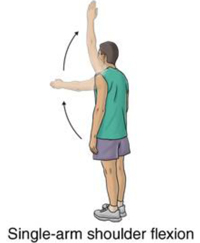
Stand with your injured arm hanging down at your side. Keeping your arm straight, bring your arm forward and up toward the ceiling. Hold this position for 5 seconds. Do 2 sets of 8 to 12. As this exercise becomes easier, add a weight.
  • Resisted shoulder internal rotation: Stand sideways next to a door with your injured arm closest to the door. Tie a knot in the end of the tubing and shut the knot in the door at waist level. Hold the other end of the tubing with the hand of your injured arm. Bend the elbow of your injured arm 90 degrees. Keeping your elbow in at your side, rotate your forearm across your body and then slowly back to the starting position. Make sure you keep your forearm parallel to the floor. Do 2 sets of 8 to 12.
  • Resisted shoulder external rotation: Stand sideways next to a door with your injured arm farther from the door. Tie a knot in the end of the tubing and shut the knot in the door at waist level. Hold the other end of the tubing with the hand of your injured arm. Rest the hand of your injured arm across your stomach. Keeping your elbow in at your side, rotate your arm outward and away from your waist. Slowly return your arm to the starting position. Make sure you keep your elbow bent 90 degrees and your forearm parallel to the floor. Repeat 10 times. Build up to 2 sets of 15.
  • Side-lying external rotation: 
    Lie on your uninjured side with your injured arm at your side and your elbow bent 90 degrees. Keeping your elbow against your side, raise your forearm toward the ceiling and hold for 2 seconds. Slowly lower your arm. Do 2 sets of 15. You can start doing this exercise holding a soup can or light weight and gradually increase the weight as long as there is no pain.
  • Sleeper stretch: 
    Lie on your injured side with your hips and knees flexed and your arm straight out in front of you. Bend the elbow on your injured side to a right angle so that your fingers are pointing toward the ceiling. Then use your other hand to gently push your arm down toward the floor. Keep your shoulder blades lightly squeezed together as you do this exercise. Hold the stretch for 30 seconds. Repeat 3 times.
BICEPS TENDON STRENGTHENING EXERCISES: