Sunday 27 May 2018

Positioning for stroke patients

Introduction:

The aim of positioning the patient is to try to promote optimal recovery by modulating muscle tone, providing appropriate sensory information, increasing spatial awareness and prevention of complications such as pressure sores, contracture, pain, respiratory problems and assist safer eating.
Correct positioning can help to reduce the risk of;
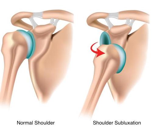
AspirationContracture Pressure AreasShoulder Pain Swelling of the Extremities 

Image result for positioning for stroke patients in bed

Aims of positioning:

Normalise Tone or Decrease Abnormal influence on the Body
Maintain Skeletal Alignment
Prevent, Accommodate or Correct Skeletal Deformity
Provide Stable Base of Support
Promote Increased Tolerance of Desired Position
Increased Stimulation to Affected Side
Increased Spatial Awareness
Promote Patient Comfort
Facilitate Normal Movement Patterns
Control Abnormal Movement Patterns
Manage Pressure
Decrease Fatigue
Enhance Autonomic Nervous System Function (Cardiac, Digestive and Respiratory Runction)
Facilitate Maximum Function
Improved Ability to Interact with the Environment

Types of positioning:

1Sitting in a Chair or Sitting in a Wheelchair:Image result for Sitting in a Chair or Sitting in a Wheelchair

It is vital that as soon as the person is capable of sitting out that they are facilitated to do so. Sitting out is essential to build up tolerance; provide maximum stimulation; give a sense of normality.

Head over PelvisHips at 90 degreesKnees at 90 degreesSlight extension of lumber regionFeet in neutral position and supportedWeight evenly distributed between both buttocksArm should be protracted forward and supported

2. Side lying on the unaffected side:

The stroke arm should be well forward, keeping the elbow straight and supported on a pillow.
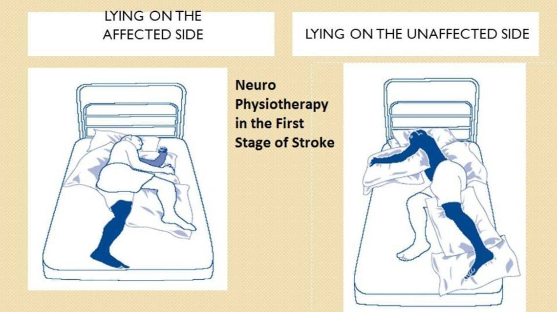
The stroke leg should be brought far enough in front of the body to prevent the patient rolling on to the back, the knee bent and leg supported on a pillow.
A small pillow can then be placed under the patient's waist to maintain the the line of the spine.
When lying on the side position, the patient should have two pillows only under the head. 
Image result for Side lying on the unaffected side

3. Side lying on the affected side:

This should always be encouraged with the stroke shoulder well forward so that the body weight is supported on the flat of the shoulder blade and not on the point of the shoulder.
One or two pillows for head
Place the stroke leg with the thigh so that it is in line with the trunk, and bend the knee slightly.
The unaffected leg should be brought forward and placed with the knee bent on a pillow in front of the affected leg for comfort. This prevents the patient rolling onto his back.
Lastly, bend the head forward a little.  

4.Lying supine:

This is the position most likely to encourage spasticity, but some patients do like to lie on their back for a while and it will be required for some treatments.Image result for Lying supine
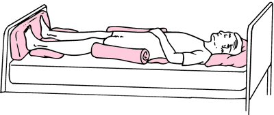
Place two pillows under the patient's head and help him/her bend their head slightly towards their unaffected shoulder and gently turn their head towards their stroke side but do not uses force.
A small pillow is placed under the buttock of the stroke side and should extend just to the knee, this will relax the leg and prevent it turning out at the hip.
A pillow is placed under the stroke arm which is kept straight at the elbow and if possible, the palms of the hand facing upwards.
The bed must be the correct height to promote independence and safety for the patient, family and health care workers. 

5. Sitting up in bed:

Sitting in bed is desirable for short periods only

Image result for Sitting up in bed
Must be upright and well supported with pillows
Consider extra support using pillows under arms or knees

Sitting versus lying:

When seated, nearly half of the body weight is supported on 8% of the sitting areas at or near the ischial tuberosities.
Therefore, interface pressures are much higher in sitting than lying

Distribution of weight when seated normally:

Buttocks & Thighs 75%
Feet flat on floor 19%
Back 4%
Arms 2%
Total 100%

Pressure relief:


 A person who has had a stroke may be susceptible to developing pressure sores 
Assess the person and decide on an appropriate cushion use in order to
1.Prevent further skin breakdown,
2.To assist with healing
3.To facilitate the patient to sit out as much as possible

Tuesday 22 May 2018

Physiotherapy Management of shoulder pain in patients with stroke

Management of shoulder pain in patients with stroke:


Shoulder pain affects from 16% to 72% of patients after a cerebrovascular accident. Hemiplegic shoulder pain causes considerable distress and reduced activity and can markedly hinder rehabilitation. The aetiology of hemiplegic shoulder pain is probably multifactorial. The ideal management of hemiplegic stroke pain is prevention. For prophylaxis to be effective, it must begin immediately after the stroke. Awareness of potential injuries to the shoulder joint reduces the frequency of shoulder pain after stroke. The multidisciplinary team, patients, and carers should be provided with instructions on how to avoid injuries to the affected limb. Foam supports or shoulder strapping may be used to prevent shoulder pain. Overarm slings should be avoided. Treatment of shoulder pain after stroke should start with simple analgesics. If shoulder pain persists, treatment should include high intensity transcutaneous electrical nerve stimulation or functional electrical stimulation. Intra-articular steroid injections may be used in resistant cases. 

Image result for management of shoulder pain in patients with stroke


Occurence:
Shoulder pain is a common complication after a cerebrovascular accident. From 16% to 72% of stroke patients develop hemiplegic shoulder pain. It may occur in up to 80% of stroke patients who have little or no voluntary movement of the affected upper limb.
Hemiplegic shoulder pain has been shown to affect stroke outcome in a negative way. It interferes with recovery after a stroke: it can cause considerable distress and reduced activity and can markedly hinder rehabilitation. Royet al demonstrated that the presence of hemiplegic shoulder pain is strongly associated with prolonged hospital stay and poor recovery of arm function in the first 12 weeks after stroke.

Image result for prevention of shoulder pain in patients with stroke
The cause of hemiplegic shoulder pain is the subject of considerable controversy. The following processes have all been postulated as causes of a painful hemiplegic shoulder: glenohumeral subluxation, spasticity of shoulder muscles, impingement, soft tissue trauma, rotator cuff tears, glenohumeral capsulitis, bicipital tendinitis, and shoulder hand syndrome.Traction neuropathy of the brachial plexus may also play a part. Unusual patterns of motor recovery or spasticity or unusually severe focal atrophy may suggest brachial plexus injury. Poor handling of a hemiplegic limb may exacerbate a pre-existing condition such as osteoarthritis. Thus, pre-morbid disease of the shoulder may predispose to hemiplegic shoulder pain. Stroke patients may suffer from pain that is caused by the stroke itself (central post-stroke pain). The role of central post-stroke pain in the aetiology of hemiplegic shoulder pain is uncertain. Abnormal tone (both spasticity and flaccidity) has been suggested as an aetiological factor in hemiplegic shoulder pain. However, clinical observations suggest that shoulder pain does not occur until spasticity develops. Most authorities agree that the aetiology of hemiplegic shoulder pain is probably multifactorial.

Prevention:

The ideal management of hemiplegic stroke pain is to prevent it happening in the first place. Various strategies have been employed in the prophylaxis of hemiplegic shoulder pain. For prophylaxis to be effective, it must be begin immediately after the stroke. Once the patient has pain, resultant anxiety and overprotection will follow.

HANDLING

Poor handling and positioning of the affected upper limb in stroke patients contribute toward shoulder pain.
The mobility of the recovering stroke patient is dependent on the assistance of nurses, therapists, doctors, other ancillary staff, and family members. It is also dependent on his/her own efforts. Handling, positioning, and transferring on a day-to-day basis can exert great stress on the vulnerable shoulder. The problem may be exacerbated by the patient's sensory and perceptual deficits. There has been concern that trauma to the constituent components of the shoulder joint may be caused by poor handling of the patient's affected arm.
Wanklyn et al studied the prevalence of hemiplegic shoulder pain and associated factors in patients with stroke. Sixty three per cent of the patients developed hemiplegic shoulder pain in the first six months after their stroke. Patients who needed help with transfers were more likely to develop hemiplegic shoulder pain. Certainly, patients with markedly decreased voluntary movement after a cerebrovascular accident frequently experience shoulder joint malalignment or subluxation in the early stages of recovery.
Careful positioning and handling of the limb are thought to prevent hemiplegic shoulder pain, but there is a range of opinions about how correct limb positioning is best achieved.
Braus et al investigated the efficacy of an information and education programme in the prevention of hemiplegic shoulder pain. All members of the diagnostic and therapeutic team as well as patients and their family were provided with instructions on how to avoid injuries to the affected limb. The investigators found that awareness of potential injuries to the structures of the shoulder joint reduced the frequency of shoulder pain from 27% to 8%.
Fitzgerald-Finch et al advocated the use of the Australian lift when handling these patients: they felt it to be of value as the weight of the patient is taken on the shoulders of the carer and the patient's shoulder is protected.

STRAPPING

Glenohumeral joint subluxation may be a contributing factor in the development of shoulder pain in this group of patients. Shai et alhypothesised that earlier radiological diagnosis of subluxation might enable more effective prevention than if it is delayed. However, this has not been proved. Despite this, a variety of slings have been designed to try to correct subluxation and pain in stroke patients with hemiplegia. Not all such devices have been successful: supportive devices developed by Buccholtz Moodie et al and Williams et al were not proved to be effective in correcting subluxation of the shoulder.Image result for prevention of shoulder pain in patients with stroke
Physiotherapists have employed various forms of strapping designed for shoulder pain or subluxation after a cerebrovascular accident. Unfortunately, the effectiveness of many of these strapping methods remains largely unproved. Ancliffe undertook a pilot study to determine the effectiveness of a strapping technique to prevent shoulder pain after a stroke. The pilot study demonstrated that strapping the hemiplegic shoulder delayed the onset of shoulder pain. In patients with subluxation and shoulder pain, use of a Varney brace has been reported to be successful: patients become asymptomatic within seven days.
Image result for strapping of shoulder pain in patients with stroke
External support can be discontinued when muscle tone around the glenohumeral joint is sufficient to prevent subluxation. An exercise programme should always accompany the use of a sling.
However, a number of authors have reported that slings may hold the limb in a poor position that is likely to cause soft tissue contracture and have an adverse effect on symmetry, balance, and body image.

PHYSIOTHERAPY

Some studies have noted that passive abduction of the hemiplegic arm can result in rotator cuff injury: this in turn causes shoulder pain.
However, therapeutic range of motion exercises done by the patients can involve passive abduction of the arm. Kumar et alanalysed the occurrence of pain in patients receiving three different rehabilitation exercise programs: range of motion by the therapist, use of a skateboard, and use of an overhead pulley. They found that patients who used the overhead pulley had the highest risk of developing shoulder pain and concluded that use of the pulley should be avoided during stroke rehabilitation.
Image result for physiotherapy for shoulder pain in patients with stroke
If impingement during range of motion exercises is determined to be the cause of hemiplegic shoulder pain, the amplitude of passive movement should be kept within the pain-free range. Caldwellet al reported that pain subsided in 43% of patients with hemiplegic shoulder pain when the amplitude of passive range of motion was reduced.
Increase in the prevalence of shoulder pain in the first weeks after discharge in patients who did not continue to exercise properly.

Treatment

Radiological investigations should exclude dislocation or fracture of the shoulder before further management is instigated. Various treatments have been suggested as being beneficial in shoulder pain after stroke: these include physiotherapy, localised cooling, infrared, ultrasound, and intra-articular injections of steroids and local anaesthetics. Until recently there has been a shortage of prospective controlled clinical trials.

PHYSIOTHERAPY

Physiotherapy has been used in the treatment of hemiplegic shoulder pain. There are two major approaches to therapy in this field: those that focus on the problem as a localised mechanical one; and those that view the problem as a neurological one. Local treatments used have included heat and cold therapy. Slings and shoulder supports have also been used. Positioning is also considered important by many authors. Other physiotherapy approaches include those of Bobath, Brunnstrom, and proprioceptive neuromuscular facilitation. Until recently, the evidence for the effectiveness of these methods of physiotherapy has been poor. Partridge examined the effectiveness of two methods of physiotherapy in the treatment of hemiplegic shoulder pain: cryotherapy or the Bobath approach. The cryotherapy approach involved the application of ice to the affected shoulder. The Bobath approach is a neurologically based holistic approach that is frequently used in the UK.
There were no significant differences between the two treatments in terms of severity of pain at rest or on movement or for reported distress. However the proportion of patients who reported no pain after treatment was greater in those who received the Bobath approach.

TRANSCUTANEOUS ELECTRICAL NERVE STIMULATION


Leandri et al evaluated the effectiveness of high intensity versus low intensity transcutaneous electrical nerve stimulation (TENS) versus placebo for patients with hemiplegic shoulder pain. Low intensity TENS involves electrical stimulation just above the level of the skin sensory threshold. High intensity TENS is sufficient to elicit muscle contraction and an almost painful sensation. The investigators found that patients who received high intensity TENS had significant improvements in passive range of motion for flexion, extension, abduction, and external rotation at the shoulder. The patients who received high intensity TENS also reported very satisfactory pain relief.

FUNCTIONAL ELECTRICAL STIMULATION

There are have been a number of studies of the effectiveness of functional electrical stimulation (FES) in shoulder pain after stroke.
Faghri et al studied the effects of a FES treatment programme designed to prevent glenohumeral joint stretching and subsequent subluxation and shoulder pain in stroke patients. They demonstrated a beneficial effect on subluxation and improvement in other parameters such as pain, range of motion, and arm function.
Chantraine et al completed a long term controlled study of the use of FES in hemiplegic stroke patients diagnosed with a subluxed and painful shoulder. They found that a 24 month FES programme was effective in reducing the severity of subluxation and pain and may have facilitated recovery of shoulder function in these patients.
As an extremely mobile joint, the shoulder sacrifices stability for mobility.  Basmajian determined through electromyographic studies that the supraspinatus, and to a lesser extent the posterior deltoid muscles, played a key part in maintaining glenohumeral alignment. Chaco and Wolf also demonstrated the importance of the supraspinatus muscle in preventing downward subluxation of the humerus. Two studies have investigated the application of electrical stimulation to the supraspinatus and posterior deltoid muscles: Baker and Parker demonstrated the beneficial effects of FES in stroke patients with a chronic shoulder dislocation. However, patients deteriorated after withdrawal from treatment though not back to pre-treatment levels. Linn et al carried out a prospective randomised study to determine the efficacy of FES in the prevention of shoulder subluxation in stroke patients. They found that FES does prevent shoulder subluxation, but this effect was not maintained after the withdrawal of treatment.

Thursday 17 May 2018

Neural Developmental Therapy and its importance in Neuro Rehabilitation

                               Neurodevelopmental treatment (NDT) is a hands-on treatment approach used by physical therapists, occupational therapists, and speech-language pathologists. ... Without NDT interventions, the patient likely will develop a limited set of movement patterns that he or she will apply to nearly all tasks.

                               Before one hundred years before, If someone having delayed milestone , cerebral palsy, or some other neuro-anomalies kids, they were thinking that there is no other chance to rehabilitate them in a  correct way. But this century, we developed in many ways in rehabilitation approach too,along with other scientific growth. One of the most important Therapy to rehabilitate these children is NDT, we can apply this approach to stroke patient also to get speedy recovery.

                              Neuro developmental therapy is a non-invasive programme of movements which aims to promote development of the nervous system and the inhibition of primitive reflexes.  By incorporating movements which are used naturally by a baby and a young child, the nervous system is gently encouraged to mature and become more open to learning.  

                              During the Therapy, your child will have a set of specific movements which are performed every day for approximately 5-10 minutes.  Each programme is specifically designed for the needs of each child and will evolve as the programme progresses.  Both you and your child are guided by your therapist through the full programme and she/he will review progress with you both at regular intervals. 


CONCEPT OF NDT:


                             NDT is an approach to neurological physio rehabilitation that is applied in patient assessment and treatment (such as with adults after cerebrovascular accident (stroke), or children with  cerebral palsy ). The goal of applying the Bobath concept is to promote motor learning for efficient motor control in various environments, thereby improving participation and function. This is done through specific patient handling skills to guide patients through initiation and completion of intended tasks.This approach to neurological rehabilitation is multidisciplinary, primarily involving physiotherapists, occupational therapists and speech and language therapists. In the United States, the Bobath concept is also known as 'neuro-developmental treatment' (NDT).

Types of NDT:

1. Bobath approach is a problem-solving neurodevelopmental(NDT) approach for assessment and treatment of individuals with cerebral palsy and other allied neurological conditions.It is named after Berta Bobath, a physiotherapist, and her husband Karel, a psychiatrist/neurophysiologist. 

2. The Brunnstrom approach is a type of physiotherapy treatment used with patients with movement problems following damage to the brain and spinal cord, (central nervous system/ CNS).

3. The Rood Approach for the treatment of central nervous system disorders was developed by Margaret Rood in the 1950s. Rood's technique can be categorized as one of facilitation and inhibition of movement.

4. Vojta Therapy. According to Vojta, reflex locomotion is activated from the three main positions: prone, supine and side lying. To stimulate the patterns of movement, there are—as described by Vojta—ten available zones on the body and on the arms and legs.

Physiotherapists focuses while applying NDT are,


                            Firstly physiotherapists focuses on Short and long term goal settings as well as give activities to do daily activities ( bathing, carrying , sleeping positions etc). Physiotherapists are assisting to reach the milestones( rolling, sitting etc). Some activities should be functional and aim to maintain optimal muscle length, improve muscle . They are focusing as well on strength and coordination, assist with balance, stability as well as mobility.

Assistive devices such as walking aids, foot or hand orthoses, may assist with gaining independence.

Saturday 12 May 2018

Lower back pain


Low back pain (LBP) is a common disorder involving the muscles, nerves, and bones of the backPain can vary from a dull constant ache to a sudden sharp feeling. Low back pain may be classified by duration as acute (pain lasting less than 6 weeks), sub-chronic (6 to 12 weeks), or chronic (more than 12 weeks).
Back pain, one of the major musculoskeletal pain problems. Nowadays peoples are mostly affected by this condition, due to our lifestyle changes, loss of physical activity, obesity, eating fast food items, etc..
All of us who treat patients with low back pain have first-hand experience of patients with a long history of chronic pain and disability. Many have had multiple treatments from alternative therapists, physiotherapists, drug therapy, talking therapies, injection therapies and surgery. 

Back pain described as “simply a mechanical disturbance of the musculoskeletal structures or function of the back. We cannot diagnose any specific pathology. We cannot even localise the source of most soft tissue pain. Some doctors and therapists do claim to be able to diagnose the site and nature of the lesion in many patients.

Nowadays many more peoples are struggling with LBP. Due to our mechanical life , our working ergonomics, stress and depression, osteoporosis, ageing process etc...

The kind of back pain that follows heavy lifting or exercising too hard is often caused by muscle strain. But sometimes back pain can be related to a disc that bulges or ruptures. If a bulging or ruptured disc presses on the sciatic nerve, pain may run from the buttock down one leg. This is called sciatica.

Kidney pain and back pain can be difficult to distinguish, but kidney pain is usually deeper and higher in the and back located under the ribs while the muscle pain with common back injury tends to be lower in the backCauses of kidney pain are mainly urinary tract infections and kidney stones

Causes for severe back pain:

Acute low back pain is most often caused by a sudden injury to the muscles and ligaments supporting the back. The pain may be caused by muscle spasms or a strain or tear in the muscles and ligaments. Causes of sudden low back paininclude: Compression fractures to the spine from osteoporosis.

In addition, symptoms of lower back pain are usually described by type of onset and duration:
  • Acute pain. This type of pain typically comes on suddenly and lasts for a few days or weeks, and is considered a normal response of the body to injury or tissue damage. The pain gradually subsides as the body heals.
  • Subacute low back pain. Lasting between 6 weeks and 3 months, this type of pain is usually mechanical in nature (such as a muscle strain or joint pain) but is prolonged. At this point, a medical workup may be considered, and is advisable if the pain is severe and limits one’s ability to participate in activities of daily living, sleeping, and working.
  • Chronic back pain. Usually defined as lower back pain that lasts over 3 months, this type of pain is usually severe, does not respond to initial treatments, and requires a thorough medical workup to determine the exact source of the pain.

Types of Low Back Pain

There are many ways to categorize low back pain – two common types include:
  • Mechanical painBy far the most common cause of lower back pain, mechanical pain (axial pain) is pain primarily from the muscles, ligaments, joints (facet joints, sacroiliac joints), or bones in and around the spine. This type of pain tends to be localized to the lower back, buttocks, and sometimes the top of the legs. It is usually influenced by loading the spine and may feel different based on motion (forward/backward/twisting), activity, standing, sitting, or resting.
  • Radicular pain. This type of pain can occur if a spinal nerve root becomes impinged or inflamed. Radicular pain may follow a nerve root pattern or dermatome down into the buttock and/or leg. Its specific sensation is sharp, electric, burning-type pain and can be associated with numbness or weakness . It is typically felt on only one side of the body.

Causes of Chronic Lower Back Pain

Pain is considered chronic once it lasts for more than three months and exceeds the body’s natural healing process. Chronic pain in the low back often involves a disc problem, a joint problem, and/or an irritated nerve root. Common causes include:
Lumbar herniated disc. The jelly-like center of a lumbar disc can break through the tough outer layer and irritate a nearby nerve root. The herniated portion of the disc is full of proteins that cause inflammation when they reach a nerve root, and inflammation as well as nerve compression cause nerve root pain. The disc wall is also richly supplied by nerve fibers, and a tear through the wall can cause severe pain.

Degenerative disc disease. At birth, intervertebral discs are full of water and at their healthiest. As people age over time, discs lose hydration and wear down. As the disc loses hydration, it cannot resist forces as well, and transfers force to the disc wall that may develop tears and cause pain or weakening that can lead to a herniation. The disc can also collapse and contribute to stenosis.

Facet joint dysfunction. There are two facet joints behind each disc at each motion segment in the lumbar spine. These joints have cartilage between the bones and are surrounded by a capsular ligament, which is richly innervated by nerves. These joints can be painful by themselves, or in conjunction with disc pain.

Sacroiliac joint dysfunction. The sacroiliac joint connects the sacrum at the bottom of the spine to each side of the pelvis. It is a strong, low-motion joint that primarily absorbs shock and tension between the upper body and the lower body. The sacroiliac joint can become painful if it becomes inflamed (sacroilitis) or if there is too much or too little motion of the joint.

Spinal stenosis. This condition causes pain through narrowing of the spinal canal where the nerve roots are located. The narrowing can be central, forminal, or both, and can be at a single level or multiple levels in the lower back.

Spondylolisthesis. This condition occurs when one vertebra slips over the adjacent one. There are 5 types of spondylolisthesis but the most common are secondary to a defect or fracture of the pars (between the facet joints) or mechanical instability of the facet joints (degenerative). The pain can be caused by instability (back) or compression of the nerves (leg).

Osteoarthritis. This condition results from wear and tear of the disc and facet joints. It causes pain, inflammation, instability, and stenosis to a variable degree, and can occur at a single level or multiple levels of the lower spine. Spinal osteoarthritis is associated with aging and is slowly progressive. It is also referred to as spondylosis or degenerative joint disease.

Deformity. Curvature of the spine can include scoliosis or kyphosis. The deformity may be associated with lower back pain if it leads to the breakdown of the discs, facet joints, sacroiliac joints or stenosis.

Trauma. Acute fractures or dislocations of the spine can lead to pain. Lower back pain that develops after a trauma, such as a motor vehicle accident or a fall, should be medically evaluated.
Compression fracture. A fracture that occurs in the cylindrical vertebra, in which the bone essentially caves in on itself, can cause sudden pain. This type of fracture is most common due to weak bones, such as from osteoporosis, and is more common in older people.

Common Symptoms of Lower Back Problems

Specifically identifying and describing symptoms can help lead to a more accurate diagnosis and effective treatment plan.
Low back pain is typically characterized by a combination of the following symptoms:
Dull, aching pain. Pain that remains within the low back (axial pain) is usually described as dull and aching rather than burning, stinging, or sharp. This kind of pain can be accompanied by mild or severe muscle spasms, limited mobility, and aches in the hips and pelvis.
Pain that travels to the buttocks, legs, and feet. Sometimes low back pain includes a sharp, stinging, tingling or numb sensation that moves down the thighs and into the low legs and feet, also called sciatica. Sciatica is caused by irritation of the sciatic nerve, and is usually only felt on one side of the body.
Pain that is worse after prolonged sitting. Sitting puts pressure on the discs, causing low back pain to worsen after sitting for long periods of time. Walking and stretching can alleviate low back pain quickly, but returning to a sitting position may cause symptoms to return.
Pain that feels better when changing positions. Depending on the underlying cause of pain, some positions will be more comfortable than others. For example, with spinal stenosis walking normally may be difficult and painful, but leaning forward onto something, such as a shopping cart, may reduce pain. How symptoms change with shifting positions can help identify the source of pain.

Pain that is worse after waking up and better after moving around. Many who experience low back pain report symptoms that are worse first thing in the morning. After getting up and moving around, however, symptoms are relieved. Pain in the morning is due to stiffness caused by long periods of rest, decreased blood flow with sleep, and possibly the quality of mattress and pillows used. 

Low Back Pain Symptoms by Location

The body’s largest vertebrae are found in the lumbar spine, supporting most of the weight of the upper body. These vertebrae are highly susceptible to degeneration and injury, and an injury at one spinal level can cause a specific set of symptoms:
L3-L4. The L3-L4 nerve root is likely to cause shooting pain in the front of the thigh, possibly including numbness or tingling. Pain or neurological symptoms may radiate to the front of the knee, shin, and foot as well, though it is less common.
L4-L5. Pain from the L4-L5 segment typically manifests as sciatic pain in the back of the thigh, and possibly pain that reaches the calves, combined with axial low back pain.
L5-S1. Where the base of the spine connects to the sacrum there are a couple of joints that provide support and flexibility. One is the lumbosacral joint, which allows the hips to swing side to side, and the other is the sacroiliac joint, which has limited mobility and mainly absorbs shock from the upper body to the low body.
Pain from the L5-S1 segment is generally caused by problems with these joints or from a compressed nerve root. Issues with the L5-S1 segment commonly cause sciatica.
Different nerve roots are irritated depending on the structures in the back that are injured, and being able to point to the specific areas of radicular pain can help more precisely diagnose the source of low back pain.

Symptoms That Require Immediate Attention

Sometimes low back pain can signal a serious underlying medical condition. People who experience any of the following symptoms are advised to seek immediate care.
  • Loss of bladder and bowel control
  • Recent weight loss not due to lifestyle changes, such as diet and exercise
  • Fever and chills


  • Physical Exam

    The goal of a physical exam is to further narrow down possible causes of pain. A typical physical for low back pain includes some combination of the following steps:
    Palpation. A doctor will feel by hand (also called palpation) along the low back to locate any muscle spasms or tightness, areas of tenderness, or joint abnormalities.
    Neurologic exam. Diagnosis will likely include a motor exam, which involves manual movement of hip, knee and big toe extension and flexion (movement forward and backward) as well as ankle movement. A sensory exam will likely include testing the patient’s reaction to light touch, a pin prick, or other senses in the lower trunk, buttock and legs.
    Range of motion test. The patient may be asked to bend or twist in certain positions. These activities are done to look for positions that worsen or recreate pain, and to see if certain movements are limited by discomfort.
    Reflex test. The patient’s reflexes in the legs will be checked to evaluate weakened reflexes and decreased muscle strength. If reflexes are diminished, a nerve root might not be responding as it should.
    Leg raise test. The patient is asked to lay on the back and raise one leg as high and as straight as possible. If this leg raise test recreates low back pain, a herniated disc might be suspected.
    Obtaining an accurate diagnosis that identifies the underlying cause of the pain, and doesn’t just correlate to the symptoms, is important in guiding treatment.
    Lower back pain
    Many structures in the low back can cause pain, making the source sometimes difficult to identify.
    As a foundation of the diagnostic process, the patient provides a detailed description of symptoms and medical history. From this information, a doctor will usually have a general idea of the source of the patient’s pai

    Patient History

    Before starting a physical exam, the patient will be asked to provide information regarding symptoms and medical history. Inquiries typically include:
    Information about current symptoms. Is the pain better or worse at certain times of day, such as waking up or after work? How far does the pain spread? Are there other symptoms at the same time, such as weakness or numbness? What does the pain feel like—achy, sharp, tight, dull, hot, stinging?
    Activity level. Does the person lead a generally more active or sedentary lifestyle? For example, does work require sitting at a desk or standing at an assembly line for long periods of time? How often does the person exercise?
    Sleep habits. As a general rule, how many hours of sleep does the patient get? What sleep position is preferred? What kind of and/or quality of mattress and pillow does the patient use?
    Posture. What kind of posture feels comfortable or uncomfortable? Does the patient typically sit upright or slouch?

    Injuries. Has the person had any recent injuries? Has there been an injury in the past that might be relevant now?
    Answers to these questions provide a doctor with a fuller picture of the patient’s daily life, indicating more specific possibilities for low back pain. A medical history is often the most powerful tool for finding a diagnosis.

    Physical Exam

    The goal of a physical exam is to further narrow down possible causes of pain. A typical physical for low back pain includes some combination of the following steps:
    Palpation. A doctor will feel by hand (also called palpation) along the low back to locate any muscle spasms or tightness, areas of tenderness, or joint abnormalities.
    Neurologic exam. Diagnosis will likely include a motor exam, which involves manual movement of hip, knee and big toe extension and flexion (movement forward and backward) as well as ankle movement. A sensory exam will likely include testing the patient’s reaction to light touch, a pin prick, or other senses in the lower trunk, buttock and legs.
    Range of motion test. The patient may be asked to bend or twist in certain positions. These activities are done to look for positions that worsen or recreate pain, and to see if certain movements are limited by discomfort.
    Reflex test. The patient’s reflexes in the legs will be checked to evaluate weakened reflexes and decreased muscle strength. If reflexes are diminished, a nerve root might not be responding as it should.
    Leg raise test. The patient is asked to lay on the back and raise one leg as high and as straight as possible. If this leg raise test recreates low back pain, a herniated disc might be suspected.
    Usually, a doctor is able to diagnose low back pain based on the information gleaned from a medical history and a physical exam, and further testing is not needed
    An imaging scan is sometimes needed to gain more information on the cause of a patient’s pain. An imaging test may be indicated if the patient’s pain is severe, not relieved within two or three months, and does not get better with nonsurgical treatments.
    Common imaging tests include:
    X-rays are used to look at the bones of the spine. They show abnormalities, such as arthritis, fractures, Bone spurs, or tumors.
    A CT scan/Myelogram provides a cross-sectioned image of the spine. In a CT SCAN(Computer Tomography) an x-ray is sent through the spine, which a computer picks up and reformats into a 3D image. This detailed image allows doctors to look closely at the spine from different angles. Sometimes a myelogram is performed in tandem with a CT scan, in which dye is injected around nerve roots to highlight spinal structures, giving the image more clarity.
    An MRI, or Magnetic Resonance Imaging scan, provides a detailed image of spinal structures without using the radiation required with x-rays. An MRI can detect abnormalities with soft tissues, such as muscles, ligaments, and intervertebral discs. An MRI might also be used to locate misalignments or joint overgrowth in the spine.
    Injection studies are fluoroscopic-directed injections of local anesthetic and steroid medication into specific anatomic structures. They are helpful in confirming the source of the pain. They are used in diagnosis, in conjunction with rehabilitation, and are considered predictive of surgical outcomes.