Thursday, 15 February 2018

QUADRICEPS TENDINITIS- REHABILITATION

Alignment or overuse problems of the knee structures can lead to strain, irritation, and/or injury of the quadricepsmuscle and tendon. This produces pain, weakness, and swelling of the knee joint.These problems can affect people of all ages but the majority of patients with overuse injuries of the knee (and specifically quadriceps tendonitis) are involved in soccer, volleyball, or running activities.
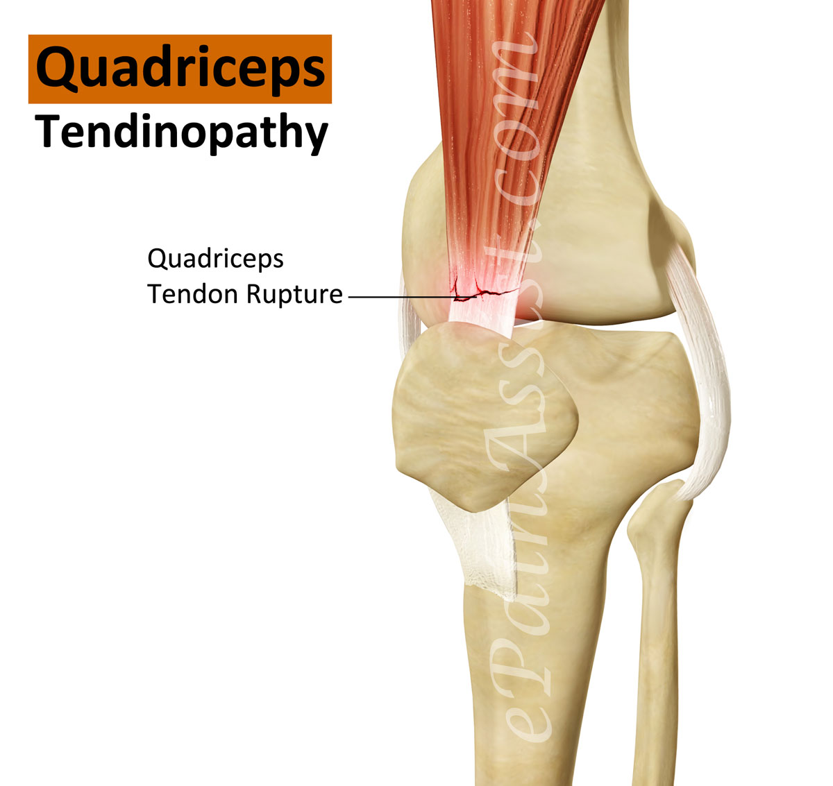
Image result for eXERCISES FOR Quadriceps tendonitisUsually , The term tendinopathy is used to cover most tendon injuries. The insertion of a muscle is its lower attachment point. The quadriceps muscles insert at the top of the patella (kneecap). This injury is an overuse injury, as the pain tends to develop gradually over a period of time, rather than at a specific point.

For a first time injury, recovery may take 2 to 3 months. A longer term chronic, recurring injury may need 4 to 6 months. Gradually return to sports specific training. Again depending on severity this may be days, it may be weeks.


Structure of Quadriceps muscle:

The large quadriceps muscle ends in a tendon that inserts into the tibial tubercle, a bony bump at the top of the tibia (shin bone) just below the patella. The tendon together with the patella is called the quadriceps mechanism. Though we think of it as a single device, the quadriceps mechanism has two separate tendons, the quadriceps tendon on top of the patella and the patellar tendon below the patella. Tightening up the quadriceps muscles places a pull on the tendons of the quadriceps mechanism. This action causes the knee to straighten. The patella acts like a fulcrum to increase the force of the quadriceps muscles.

Causes:

Quadriceps tendonitis occurs most often as a result of stresses placed on the supporting structures of the knee. Running, jumping, and quick starts and stops contribute to this condition. Overuse injuries from sports activities is the most common cause but anyone can be affected, even those who do not participate in sports or recreational activities.


Extrinsic factors: 1) Inappropriate footwear 2) Training error 3) Surface area of the ground

Intrinsic factors: 1)  Flexibility, and joint laxity 2)  Malalignment of the foot, ankle, and leg.

SYMPTOMS:

Pain from quadriceps tendonitis is felt in the area at the bottom of the thigh, just above the patella.


Tenderness develops in the area of the tendon attachment above the kneecap.
May be swelling in and around the quadriceps tendon.
May feel a sense of warmth or burning pain.
Stiffness of the knee is common when you first get up in the morning

PHYSIOTHERAPY MANAGEMENT:

The initial treatment for acute quadriceps tendonitis begins by decreasing the inflammation in the knee. 

Physical therapy can help in the early stages by decreasing pain and inflammation. Your physical therapist may use ice massage, electrical stimulation, and ultrasound to limit pain and control (but not completely prevent) swelling. Some amount of inflammatory response is needed for a good healing response. Heat may be used in cases of chronic tendinosis to stimulate blood circulation and promote tissue healing.Your physician may suggest relative rest and anti-inflammatory medications, such as aspirin or ibuprofen.

Relative rest is a term used to describe a process of rest-to-recovery based on the severity of symptoms. Pain at rest means strict rest and a short time of immobilization in a splint or brace is required. When pain is no longer present at rest, then a gradual increase in activity is allowed so long as the resting pain doesn't come back.


The therapist will prescribe stretching and strengthening exercises to correct any muscle imbalances. Flexibility exercises are often designed for the thigh and calf muscles. Specific exercises are used to maximize control and strength of the quadriceps muscles. You will be shown how to ease back into jumping or running sports using good training techniques


Bracing or taping the patella can help you do exercises and activities with less pain. Therapists also design special shoe inserts, called orthotics, to improve knee alignment and function of the patella. Proper footwear for your sport is important. The therapist will advise you in this area.


Prevention of future injuries through patient education is a key component of the treatment program. This is true whether conservative care or surgical intervention is required. Modification of intrinsic and extrinsic risk factors is essential.

Taping:

Kinesiology tape is supposed to last several days, but to achieve this the skin needs to be clean and dry or the tape will peel off. Wash and dry the area thoroughly, and do not apply any oils, lotions or moisturisers


.

Step 1: Start with a strip of kinesiology tape long enough to reach from the mid-thigh to the top of the shin. With scissors, split one end of the kinesiology tape into two equal ‘tails’ by cutting lengthways down the centre of the strip. Take the split about halfway along the strip of tape. Then use the scissors to round off all three ends of the tape.

Step 2: With the knee bent at 90 degrees, apply the unsplit end of the kinesiology tape down the midpoint of the front thigh. Apply the tape without any stretch. The point where the split begins must be above the kneecap:
Step 3: Fan the split ends of the kinesiology tape around the kneecap. Bring each strip of tape down either side of the kneecap, and then around below it. Again, apply the tape with no stretch. The two split ends should cross each either in the soft area just below the kneecap:
Step 4: Finally, give the kinesiology tape a good rub with the flat of the hands to warm it up and activate the adhesive.

Exercises to improve the condition of Quadriceps Tendinitis:


Static Quads

A static quads contraction is a good beginning exercise for quads tendonitis rehab.  There is less risk of experiencing pain or aggravating the knee joint at this stage. Do this exercise by lying or half-sitting on a mat on the floor with legs stretched out in front of you. Place a rolled-up towel underneath your knee. Contract your thigh muscle such that the back of your knee pushes into the towel. Hold for approximately 10 seconds. Release and repeat 10 times for a total of three sets.

Straight Leg Raise

The straight leg raise exercise strengthens the quadriceps muscle on the front of the thigh. Do this exercise by lying on your back on a mat. Lift your leg up into the air approximately 5 to 10 inches and hold for ten seconds. Return to start and repeat 10 to 12 times for a total of three sets. 

Quads Stretch
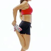
Image result for eXERCISES FOR Quadriceps tendonitis
Stretching the quadriceps muscle will help release tight structures and increase flexibility. Bend your affected leg up and grab your ankle with the hand on the same side of the body. Lift your ankle up toward your buttocks until you feel a stretch on the front of the thigh. Hold for approximately 20 to 30 seconds and repeat two to three times. Avoid bending forward at the waist during this exercise, which will take the emphasis of the stretch off the quad muscles.

Considerations for exercises:

Exercises for quadriceps tendonitis should be performed pain-free. If you are experiencing pain or an increase in pain, you may need to back off exercises or rest more. Consult with a physiotherapist who can advise you on how to treat your injury. Furthermore, your exercise program should consist of core-, gluteal- and hip-strengthening exercises to strengthen surrounding and supporting muscles and prevent muscular imbalances. Thank you.

Monday, 12 February 2018

MERITS AND DEMERITS OF CERVICAL COLLAR

Cervical/neck collars are commonly used by patients who have had a surgical intervention of the cervical spine, to immobilise the neck. It is also used for the treatment of neck pain, caused by acute trauma or chronic pain. After a whiplash injury, the neck collar can be used for both immobilisation and to reduce pain, although the value of the collar over early active mobilisations is questioned as early mobilisations can give a greater improvement in cervical range of motion and in the reduction of pain following a whiplash injury. The main goal of neck collars is to prevent or minimise motion in the cervical spine. It also keeps the head in a comfortable gravity aligned position, maintaining normal cervical lordosis.

TYPES:

Based on the materials and the hardness of the material, cervical collars can be classified into:
  1. Soft collar:
    Soft collars are made out of felt. They are cut to mould around the neck and jaw of the patient, the size being adjusted to the patient. These collars do not completely immobilise the neck however, they restrict motion and are a kinesthetic reminder for the patient to reduce neck movement. Since the collar is under the chin and supports the chin, it minimises muscle contraction needed against the gravity forces to keep the head in a normal position .This type of collar does not truly immobilise the neck, it only limits flexion and extension in the end phase.
  2. Rigid collar:
    The rigid collars are a similar design to the soft collars, but are constructed out of Plexiglas. They are easily applied and are easy to keep clean, an advantage of the plastic collar. This type of collar is also supplied in different sizes to fit the patient. These collars restrict motion in flexion and extension.They not only support the chin but also the occiput, reducing active extension, especially in the end phase. A drawback of the rigid collars is that they potentially can cause venous outflow obstruction, which may elevate intracranial pressure. If there is a clear evidence of an increased intracranial pressure, the collar should be removed or re-positioned.
    The most frequently prescribed are the Aspen, Malibu, Miami J, and Philadelphia collars. All these can be used with additional chest and head extension pieces to increase stability. Cervical collars are incorporated into rigid braces that constrain the head and chest together. Examples include the Sterno-Occipital Mandibular Immobilization Device (SOMI), Lerman Minerva and Yale types.  

Effects:


Recommendation is that a collar should be worn constantly for one week only for the reason of pain relief. After that the use of the collar should be gradually decreased. If the collar is worn for a longer period, it could have several negative effects such as: soft tissue contractures, muscular atrophy, loss of proprioception, thickening of subscapular tissues and coordination, but also psychological dependence

Comparision: 

When different types of cervical collar are compared with respect to mechanical stability (both actively and passively), all collars restrict motion to some extent. In order of least restrictive to most restrictive are: soft collar, Philadelphia collar, SOMI. although, the differences are not large. In general the collars do not provide a high level of mechanical restriction of motion and is variably between people.
The soft and rigid collar show no significant differences in movement for the most daily activities. This is because the ADL require only a small percentage of the total range of motion.Both collars can be used for people who are in less pain but need the collar to immobilize the neck and for a sense of security. In this case, the collars act primarily as proprioceptive guides to regulate the movement of the cervical spine rather than as a restraint to physically impede motion. 

Some studies show effectiveness on radiculopathy:  

Cervical collar use and rest or physiotherapy and home exercises were compared with a 'wait and see' policy for patients with cervical radiculopathy over a period of 6 weeks.
The cervical collar was semi-hard, comfortable and in six different sizes. Patients had to wear it during the first 3 weeks the whole day while also taking as much rest as possible. During the 3 last weeks, they had to decrease the time of wearing a collar a day. After 6 weeks they had to stop wearing it. Physiotherapy included exercises for mobilisation and stabilisation of the cervical spine, and reinforcing superficial and deep neck muscles. Patients also had exercises to do at home. The 'wait and see' patients were asked to continue daily activities as much as possible.
Results show that arm and neck pain were significantly reduced with the collar and physiotherapy in comparison to the wait and see policy. The results for the neck disability index show a significantly greater improvement for the collar, while physiotherapy showed the same pattern, but it wasn’t significant compared to the wait and see policy.
The cervical collar and physiotherapy decrease foraminal compression and inflammation of the nerve root by immobilisation, reducing arm and neck pain. Physiotherapy aims to regain range of motion and strength of the neck musculature, so that musculoskeletal problems are avoided. The reason for pain reduction is still unclear. It is concluded that a semi-hard cervical collar and rest, or physiotherapy and home exercises are effective for the short term (6 weeks) reduction in pain for patients with cervical radiculopathy, in comparison with wait and see policy. Primary outcome measures were VAS for neck and arm pain and the neck disability index.

Friday, 9 February 2018

PHYSIOTHERAPY FOR HAMSTRING MUSCLE STRAIN

Hamstring Tendinitis

The Hamstring tendon is the soft tissue which connects the hamstring muscle to the outer aspect of the knee. Hamstring tendinitis occurs when this tendon becomes damaged or inflamed due to excessive strain,overuse or force being placed on the tendon. 
The most common cause of hamstring tendinitis is through overuse, particularly in patients who participate in running and jumping sports. This is also common in sports such as football due to the rapid changing of speed which is undergone while running.
Whatever it is, this injury is typically seen as a result of overuse, hamstring tendinitis can also occur suddenly if the tendon becomes over stretched for example when warm-up exercises have been missed out or are inadequate for the level of activity, causing the patient to over strain the hamstring while performing running, kicking, jumping or skipping movements.

Individuals who have just started exercising or have increased their level of fitness are also at risk of sustaining this injury as they may lack strength and flexibility need for their new level of activity.

Causes

  • Overuse injury common in running and jumping activities
  • Excessive speed changing while running
  • Insufficient warm up exercise
  • Poor core strength


Hamstring Tendinitis Symptoms

Patients who suffer hamstring tendinitis are likely to feel pain in the back of the knee which gradually becomes more apparent through continued activity. This is due to the tendon becoming inflamed and swollen and in more severe cases; this pain and swelling can be felt in the thigh and calf muscles. When the injury is caused by sudden pressure or force to the tendon, pain will usually come on suddenly at the point of injury.

Patients are likely to feel aching and stiffness which becomes more apparent first thing in the morning and often the knee joint will feel a weak, making the patient unable to resume activity.

Hamstring Tendinitis Treatment

In order to treat hamstring tendinitis, rest is crucial in order to allow the injury time to heal and to prevent any further activity which could cause damage or discomfort to the injury. Applying ice to the injury is an important aspect of healing as it will reduce any swelling and inflammation as well as providing cooling pain relief to the injury. Using the PRICE method (protection, rest, ice, compression and elevation) is beneficial for the first 72 hours of the injury occurring as it will reduce swelling to the area and by keeping the injury elevated blood flow is restricted which can prevent further inflammation.
Anti-inflammatory medications can also be taken in order to reduce swelling as well as relieving pain or acheness from the injury.
Taping and strapping techniques are often used for hamstring injuries as they can help stabilize and support the area whilst relieving pressure. Some athletes find it beneficial to continue with these methods after recovery to prevent a re occurrence of the condition.
When the injury has begun healing and you are advised by a physiotherapist or doctor, it is important for the patient to undergo a rehabilitation program such as strengthening and stretching exercises which will keep the area strong and make it easier to return to sports once the injury has fully recovered. These exercises should not be carried out if the patient feels pain in the area and the advice of a physiotherapist should always be sought in order to obtain an exercise regime suitable for the individual.

Physical therapy Treatment|:

Patient should starts treatment as early as possible. The earlier the rehabilitation, the faster he/she will return to previous normal function.

Therapy takes from weeks to months depending on the condition and the history of previous injuries. 

Controlling the pain is one of the important therapy goals, this is possible through ice, electrical stimulation of the tendon and pulsed ultrasound.

Correction of pelvis misalignment has to be taken into consideration, as it might increase the tension on the hamstring muscles,and affects muscle strength. Anterior pelvic tilt is the most common misalignment and it could be easily corrected by manual or chiropractic manipulation.

Soft-tissue mobilization has to be included in the rehabilitation program. It’s very beneficial to break up the adhesions and scar tissues. A friction treatment with transverse frictions is commonly used. The therapist has to pay attention to not compress directly on the ischial tuberosity as it can irritate underlying edema. Techniques like ART (Active Release Technique) or Gastron can be included as well.

At the same time the patient may start a gradual stretching program for the hamstrings. starting with double-leg non weight bearing isometric exercises followed by single-leg closed-chain isometric and isotonic open-chain exercises.

Eccentric muscle strengthening program is a good treatment program for teninopathies , as it can normalize the thickness and structure of the tendon. It can also prepare the hamstrings for the high-force load while running.

Core strength is a considerable element for the rehabilitation of hamstring origin tendinopathy as it reduces the risk of recurrent hamstring strains..

If the ROM of the muscle is normal and pain-free, pool running and stationary biking could be put into the rehabilitation program. 

Stretching of both legs is crucial for balance. Even the antagonist hip flexor muscles should be stretched for an optimal function.

ROM can be increased by the use of ultrasound or shock wave therapy before stretching. Frequent stretching may avoid a recurrence of the injury. Some of useful Exercises are,

Standing Forward Bend With Raised Leg

Stand up straight and place the heel of one leg on a chair,  Make sure your leg is not above hip height or below knee height and that your support leg is straight, but the knee is not locked. Bend forward from your hips, keeping your back and the raised leg as straight as possible, and stop when you feel the stretch along the back of your raised thigh and knee. Don't try to touch your toes, as this will move the stretch to your back. The aim is to restrict the stretch to your hamstring muscles in the raised leg as much as you can. Hold the stretch for up to 30 seconds, then slowly return to an upright position and repeat the stretch on your other leg.

Standing Forward Bend With Crossed Legs

Stand up straight and cross one foot over the other, at your ankles, keeping both feet on the floor, says The Walking Site. Depending on how tight the backs of your legs are, this position may give you enough of a stretch down the back leg. To increase the stretch, slowly bend forward from your hips, walking your hands down a wall for stability if you need to. Only go as far as is comfortable--you should feel a tug, but no pain--and stop when your back is parallel to the floor, says The Walking Site. Keep your hands on your hips if you need to support your lower back in this position; otherwise, let your arms fall straight down so your fingers are pointing toward the floor. Hold for up to 10 seconds, then slowly return to the starting position and repeat with your legs crossed the other way.

Stretch for the Back of the Knee.

According to The Walking Site, this exercise not only stretches the backs of your knees and hamstrings, it also lengthens your calf and back muscles. Stand facing a step and position one foot so the heel is on the ground, the ball of the foot is raised, resting on the edge of the step, and your toes are pointing in the air. Your other foot should be flat on the ground. Slowly bend forward from the hips, reaching your fingers toward your raised toes. Try to keep both legs as straight as possible throughout the exercise, but only go as far as is comfortable--don't force the stretch, says The Walking Site. Hold for up to 20 seconds, then slowly and carefully return to the start position before returning the upraised foot to the ground and repeating on the other side.

Seated Stretch with Rotation

Sportsinjuryclinic.net says to sit on the floor with one leg stretched straight out in front of you and the other one bent so your foot is resting against your other inner thigh. Slightly turn the outstretched leg inward, then bend forward from your hips. You should feel the stretch up the back of the straight leg. Hold for 30 seconds, then repeat on the other leg. To move the stretch along the inner thigh, start in the same position, but slightly rotate the straight leg outward. Lean forward from the hips and hold for 30 seconds. It is more important to keep your back and outstretched leg straight than to get your chest close to your leg. Don't force the movement, but as your flexibility increases, try to bend a little deeper.

Standing Hamstring Stretch.

Stand up straight, with your legs hip-width apart and one foot slightly behind the other, says Sportsinjuryclinic.net. Keep your front leg straight, but bend your back knee and lean forward from the hips, taking care not to hunch over as you lower your torso toward your knees. You can rest your hands gently on your bent knee for support or, for an extra balance challenge, try stretching them out to your sides. Hold the stretch on each leg for about 30 seconds. If you need more of a stretch, try pointing the toes of your outstretched foot up toward the ceiling

THIS VIDEO SHOWS THAT THE STRENGTHENING EXERCISES FOR HAMSTRING TENDINITIS

Tuesday, 6 February 2018

DUCHENNE MUSCULAR DYSTROPHY

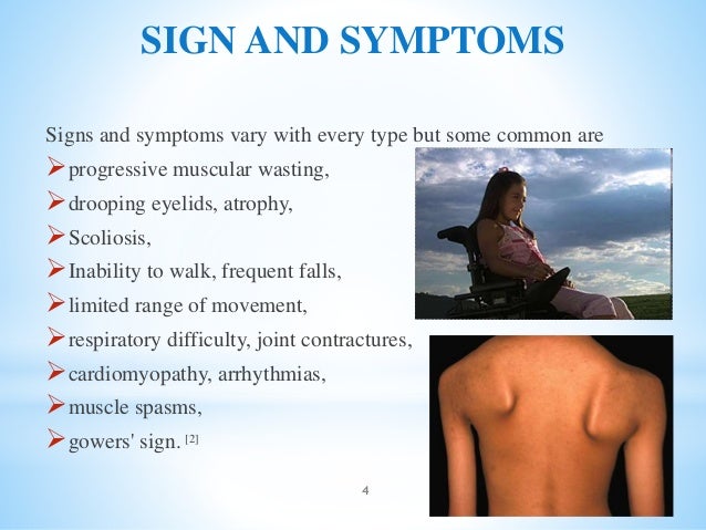
Duchene muscular dystrophy (DMD) is a genetic condition which affects the muscles, causing muscle weakness. It is a serious condition which starts in early childhood. The muscle weakness is not noticeable at birth, even though the child is born with the gene which causes it. The weakness develops gradually, usually noticeable by the age of three. Symptoms are mild at first, but become more severe as the child gets older. Duchene muscular dystrophy, the most common type, is one of more than 20 muscular dystrophies.

Image result for duchenne muscular dystrophy signs
The incidence of DMD globally is every 1/3500 male births. All types of muscular dystrophy are caused by faults in genes (the units of inheritance that parents pass on to their children) which result in progressive muscle weakness due to muscle cells breaking down and gradually becoming lost. The Duchene type affects only boys (with extremely rare exceptions) and a problem in this gene is known to result in a defect in a single important protein in muscle fibers called dystrophin.
The symptoms usually start around age 1-3 years, and may include:
Difficulty in walking, running, jumping and climbing stairs. Walking may look different with a 'waddling' type of walk. The boy may be late in starting to walk (although many children without DMD also walk late).

Image result for duchenne muscular dystrophy signs


When you pick the child up, you may feel as if he 'slips through your hands', due to looseness of the muscles around the shoulder.

Toe-walking, Frequent falls

The calf muscles may look bulky, although they are not strong.

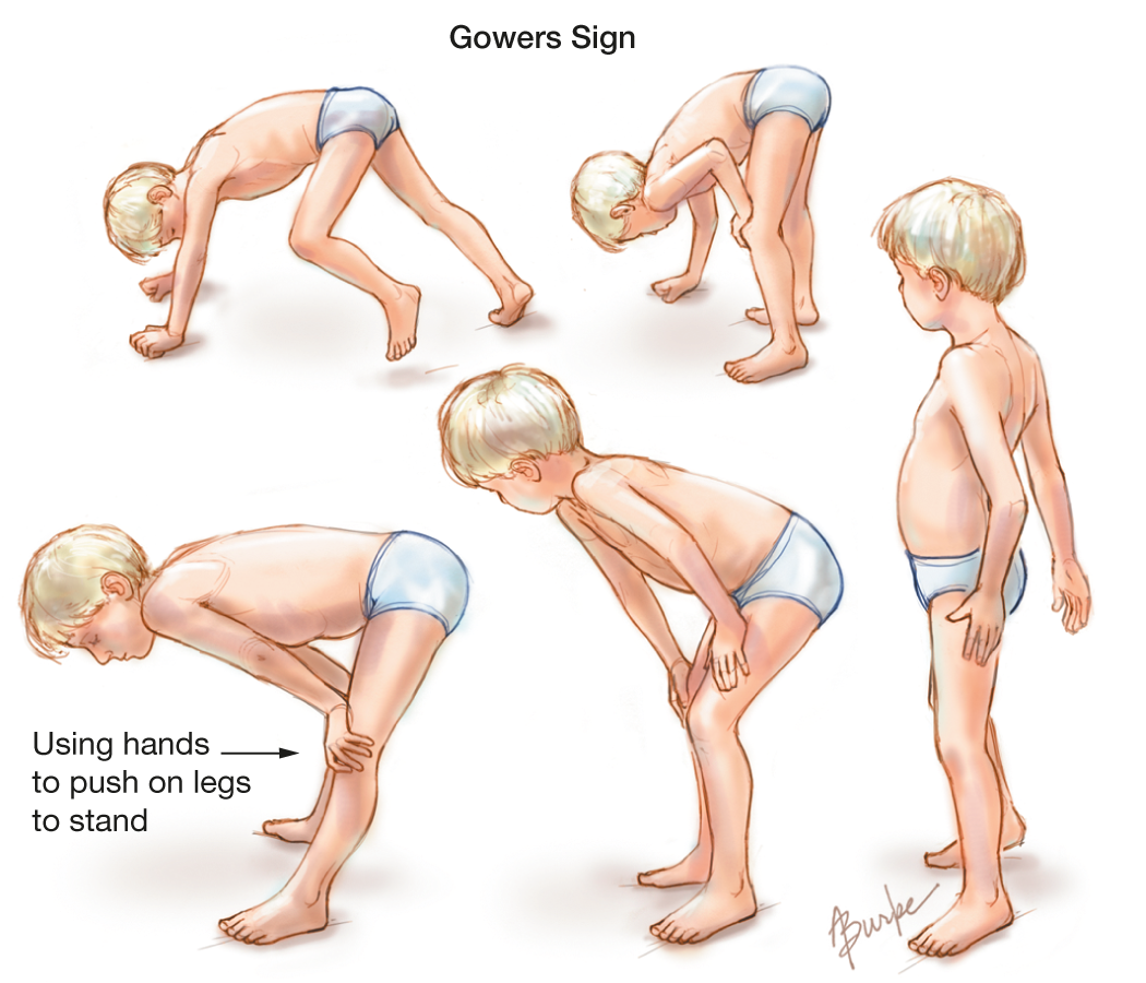
As he gets older, the child may use his hands to help him get up, looking as if he is 'climbing up his legs'. This is called 'Gower's sign'.Image result for duchenne muscular dystrophy signs

PHYSIOTHERAPY MANAGEMENT:

Image result for duchenne muscular dystrophy signs

 Physiotherapists can help slow the degression of range of motion, muscle strength, daily function, work to improve gait pattern and posture/alignment . Physiotherapy can also address the pain that the patient may be experiencing. As the patient's walking and standing abilities decline the physiotherapist may choose to implement a standing program 


Physiotherapists do stretching program with combination of passive range of motion, active range of motion, and active assisted range of motion. Regular stretching of the ankle, knee, and hip is necessary throughout the course of a patient's life. Physiotherapists will teach you, how to stretch these muscles himself.

Role of Physiotherapy in Muscular Dystrophy
The main goal of physiotherapy in patients with MD is to maintain the available function in their limbs and attain maximum possible improvement in the associated disabilities. This would help the patient attain a socially functional status so that he/she can function in the surroundings, of their own accord, bringing about a sense of independence in the patients.

Image result for duchenne muscular dystrophy PHYSIO TREATMENT


The therapeutic practices involved and their importance in the life of an MD patient are as follows:

  • Electrotherapy: A patient with MD often complains of musculoskeletal pains at various sites. Electrotherapeutic modalities like paraffin wax bath over fractured limb, TENS, IFT, Ultrasonic therapy for tender points and Contrast Bath can be done to relieve such pains.
  • Passive movement: Passive movements are a technique that involves movement of limbs by the physiotherapist, in all ranges, in a manner so as to maintain joint and muscle integrity. Long term immobilization in patients due to weakness of the muscles can render the muscle fibrotic and the joint may become stiff. To avoid the development of these co morbidities one need to maintain the integrity of structures by passive movements.
  • Active assisted movements: The MD patient has limited muscle power. Therefore, with maximum effort, they can attain only a limited range by active contraction of their muscle. To maintain this power, maximum amount activity upto the fatigue threshold should be encouraged in these patients. While the patient maintains muscle force, the physiotherapist should assist the completion of this movement in the normal biomechanical pattern. This will maintain the joint proprioception i.e. the sense of joint position in space.
  • Stretching manoeuvres: Often again due to immobility and poor power of muscles, the muscles shorten in length. The joints adapt a gravity assisted position and internal muscular forces cannot work against the external gravitational force. As a result of this joint contractures develop. To open up these joints and retain the normal muscle length, stretching is done on the joints. A sustained long duration stretch with crepe bandages or taping can be given initially and this can be toned down a bit in later stages to short duration stretches with greater repetitions given manually.
  • Joint mobilization: Due to imbalance of muscular forces, the joints often get displaced from their normal anatomical position. To guide them back so that proper weight bearing can be done on the joints without causing any harm to the associated structures, the physiotherapist passively mobilizes the bones of each joint to bring them in place.
  • Balance and Gait training: Muscular imbalances are so profound in MD that sitting and standing balance are greatly affected in the patients. So with gradual progression from kneeling to quadruped to high sitting to standing position, balance training should be given. As the balance improves the patient will be able to function better by himself/herself. With gradual degradation of power in lower extremities, the locomotion or gait is affected. So gait training involving proper training in parallel bars progressing from supported to unsupported walking should be done under the supervision of the physiotherapist.
  • Hydrotherapy: Hydrotherapy or aquatic exercises are an innate part of physiotherapy rehabilitation protocol for MD. Activities are performed in water at a warmer temperature than body. This helps in the following manner:
    • Buoyancy of water protects and braces the weak joints.
    • In water a person can feel very little of his/her own weight so this makes activities of partial weight bearing possible.
    • Adding floatation devices can assist the movements while adding high pressure water jet can help in performing mild resistance training in better muscle groups.
    • Warmer water helps in maintaining good thermo-stasis in the body and keeps the active muscle warm and hence at ease.
  • Cardiac Pacing and Breathing Exercises: As the muscles of heart and respiratory system weaken, greater chest and cardiac congestion is seen in the patients with MD. To avoid the deleterious effects of an insufficient cardiopulmonary system, one needs to keep in mind a few points.
    • Clear airway should be maintained by passive chest manipulations given by the physiotherapist.
    • The patient should be taught huffing and coughing to aid him in spitting out the chest secretions.
    • Deep breathing exercises should be taught to condition the general cardiorespiratory performance and endurance.

Saturday, 3 February 2018

ACCUPUNTURE AND PHYSIOTHERAPY

Nowadays Acupuncture is becoming more widely used and accepted treatment throughout the medical community. Many physiotherapists offer acupuncture as a treatment modality as part of physiotherapy management. Dry needling is a kind of acupuncture technique, we are using worldwide.


Acupuncture

Acupuncture techniques were used by Chinese in olden days, Acupuncture involves inserting fine needle into specific points located throughout the body to help relieve pain or have other therapeutic effects. There may be a mild aching sensation around the site of the needle, they are extremely fine and are often hardly felt.  There are many modern theories as to why acupuncture works and there has been much scientific research carried out to help prove these theories.


When physiotherapists use Acupuncture, they use disposable, single use, pre-sterilized needles of varying widths and lengths. A number of needles may be used at each treatment and these are typically left in position for up to 30 minutes before being removed depending on the response required. A needle may then be manipulated until a tingle sensation is felt (a tingling or often warm heavy sensation that can spread away from the area of the needle).
Pain relief may be immediate or develop over a few hours or days. Acupuncture often has a cumulative effect, with pain relieving affects building up over a series of treatments. There are some possible side-effects, mostly they are rare, but can include light-headedness, dizziness, tiredness, slight bruising or muscle aching after treatment. 
What Acupuncture doing in treatment?
Acupuncture may be suggested by your physio for a variety of conditions. It is often used as an adjunct in the management of:
  • Acute and chronic injuries
  • Back and neck pain
  • Muscle and joint problems
  • Nerve pain
  • Headaches and migraines
  • Sports injuries
Qualified practitioner

Physiotherapists choose to complete courses which cover trigger point dry needling only (dry needling is the use of needles for therapy of muscle pain/ tension).
It’s advisable that physiotherapists who use acupuncture and /or dry needling as a treatment with patients have completed an appropriate level of training and are engaged in ongoing professional development activities. You should feel free to ask your physiotherapist about the level of training they have completed.
Acupuncture is safe method of treatment:
Acupuncture, when provided by an appropriately trained physiotherapist is generally very safe. However with all treatments there is a small amount of risk. Your physiotherapist will discuss the risks associated with your specific treatment, and if necessary can discuss alternative treatment options with you. If you are feeling uncomfortable about a particular method of treatment, you have the right to consult another physiotherapist for a second opinion. 
It is one of the more profitable and acceptable method to overcome the pain....